
"Alternatives" to animal experiments
What does the term “alternative methods” actually mean? The term is in quote marks because it implies animal experiments could easily be replaced by another “alternative” method. However, animal experiments are ethically and especially scientifically outdated. Animal experiments are even fatal and misleading as they do not provide reliable research results translatable to humans, thus inhibiting medical progress. It should further be noted “alternative methods” are not automatically animal-free. They also include methods that only reduce the animals’ suffering or require fewer animals. More on this subject in the section on the 3R principle.
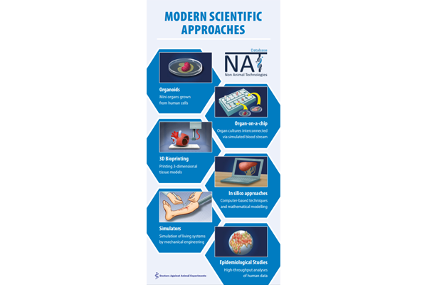
Animal-free methods using human cells or human data provide relevant results for humans, which offers a decisive advantage compared to animal experiments. The field of animal-free technologies advances rapidly and includes a wide spectrum of options, such as cell culture research, computer models, complex multi-organ chips, or epidemiological studies. They are not merely “alternatives” replacing animal experiments but highly relevant systems enabling crucial advancements in medicine and research - in contrast to animal experiments.
In the following article you will find a selection of innovative animal-free research methods. You may screen our database of non-animal technologies for specific methods and fields.
Preliminary remarks
While it is true many of the animal-free methods cannot predict how an entire organism, namely the entire human body, will respond, animal experiments cannot do that either. Animals do provide an entire organism, yet it is the wrong organism. In contrast, animal-free methods using human cells and tissues, mini organs, or multi-organ chips, combined with special computer programs based on human data, are able to provide precise, and for humans, relevant results.
- In vitro: testing systems using test tubes and painless matter like cells or tissue
- In vivo: experimenting on the living organism
- In silico: computer simulations
Why animal cells are the wrong choice
Many modern testing techniques include animal-derived material, such as cells, tissue, organs, or slaughterhouse waste. Such models are ethically unjustifiable, just like animal experiments. From a scientific point of view, it does not make sense to conduct research on animal cells or organs as they derive from the wrong organism and thus include severe errors in their results, as more and more researchers come to recognize. If, for instance, sunblock is tested on skin cells derived from mice, it cannot be predicted how the human skin in general and the different types of skin in particular will respond. Much too great are the differences in the structure of the individual skin layers of mice and humans, as well as the sensitivity of the human skin types.
Fetal calf serum
It is a positive development seeing more and more researchers work with cell or tissue cultures, thus moving away from animal experiments. The cell cultures, however, need a growth medium in order to be kept alive and grow. Therefore, it is still common practice to use fetal calf serum (FCS), as it is called. This serum is gained from the blood of unborn calves by sticking a needle directly into their hearts. The calf is drained of all the blood and dies. This procedure cannot be justified ethically. Besides, there are growth cultures scientifically superior to FCS, e.g., gained from human blood. You can find more on this here. You can find more on this here: FCS-free media >>
3R – Reduce, Refine, Replace
When dealing with “alternative methods,” one again and again comes across the term 3R. What does it mean? 3R means Reduce, Refine, Replace.
The British scientists W. Russel and R. Burch launched the concept of 3R in 1959.
Animal experiments as a method are not questioned. Rather, the idea is to improve the established system of animal experiments through Replacement, i.e., painless or less pain-sensitive systems; Reduction, i.e., reducing the number of animals; or Refinement, i.e., reducing the pain for animals, or providing more animal-friendly housing conditions. Turning away from animal experiments is not considered in this concept. Our elaborated position on the 3R concept can be found here >>
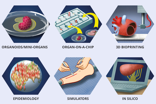
Innovative animal-free methods
Cell research
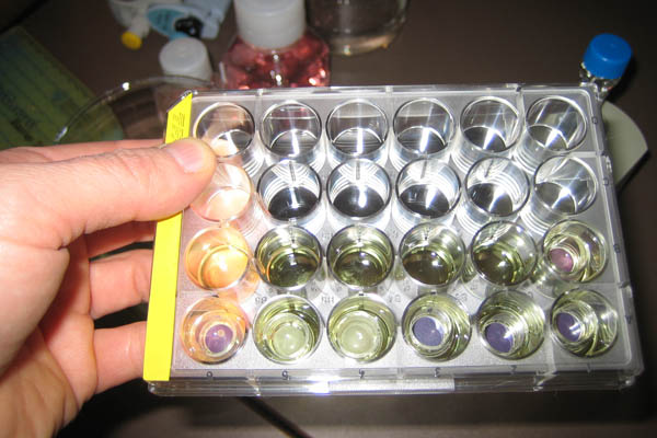
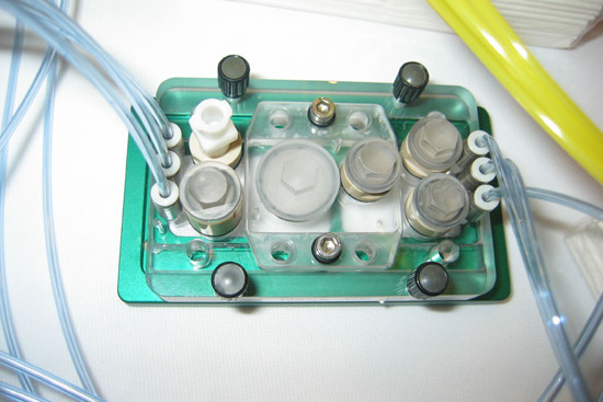
Cell cultures are grown in such well plates as these.
We distinguish between primary and continuous cell cultures. Primary cells are gained directly from the organism. Human cells - gained, for instance, from liver, skin, cartilage, or bone marrow - derive from “waste material” from clinically necessary operations, organ donations, as well as placentas and umbilical cords from births. Primary cells die after a certain period of time. Thus, cultivating them is only temporarily possible.
Cells that can be cultivated over long periods of time are known as continuous cells. They can divide infinitely and can virtually live infinitely. Tumors, for example, are typically able to do that. Nowadays, there are many thousands of different cell lines.
Thanks to most modern techniques, even complex structures of the human body can be “reconstructed” in test tubes. It is possible, for instance, to recreate human skin with its diverse layers of different cells, as well as three-dimensional heart, liver, and cartilage tissues or blood vessels.
Heart muscles cells, for example, allow us to study physiological processes and the effects of heart medication. The human eye cornea can be reconstructed with all of its layers and can be used, for instance, to test eye drops.
A system of human liver cells can be used to test new drug substances. In a comparative study, an anti-cancer substance was simultaneously tested in a clinical study on humans, rats, and the human liver cell system. The results of the tests on humans and liver cells matched. The animal experiment, in contrast, provided a misleading result (1).
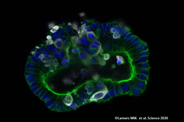
In 2010, scientists succeeded in growing organoids, as we call them, for the first time. They are three-dimensional cell cultures that realistically reflect the function of human organs. They are made out of human skin or hair-root cells. With the support of genetic engineering methods, the cells are reprogrammed into induced pluripotent stem cells and are able to produce any type of cell.
Research on cells and tissues allows us to gain valuable knowledge for humans. Critics often dismiss them as being too simple because they are unable to represent an entire organism. While animal experiments are not able do that either, animal-free research can accomplish much more than animal experiments, as will be strikingly shown in the following sections.
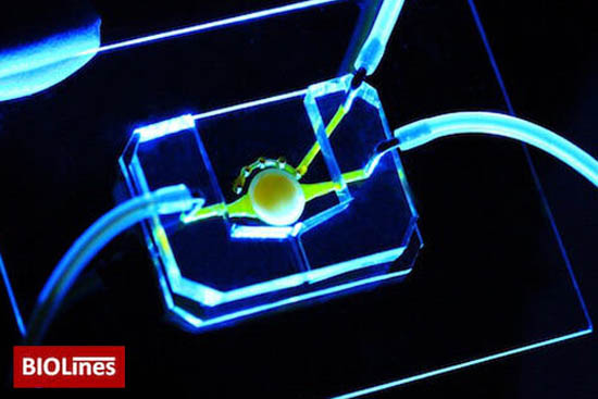
Organ-on-a-chip technologies

Mini-organs or organoids are representations of real organs and only a few millimeters in size. They are typically made out of induced pluripotent stem cells (iPSC) and put on a small plastic chip. This miniaturization allows the various tests to be automatized so that thousands of substances can be tested reliably, cost-effectively, and fast within a short period of time. In contrast to animal experiments, they can also be translated to the human body. These organ chip systems are not only advancing due to ethical reasons, sparing numerous animals death in the lab, but they are also much more effective. Furthermore, they lay the foundation for personalized medicine. In the future, a patient-on-a-chip may be generated out of a patient’s cells in order to test medication tailored toward that patient.
In the meantime, a variety of such fantastic organs-on-a-chip can be used. Here is a small selection. More about mini-organs and multi-organ-chips >>
Neuron-on-a-chip
A team of scientists at the Leibniz Institute for Analytical Sciences, the Leibniz Research Center for Working Environment and Human Factors, and TU Dortmund University have developed a nerve net on a biochip that has significant benefits over animal experiments and even conventional cell cultures. Healthy nerve cells (neurons) in the brain also try to produce extensions and get in touch with each other in a culture dish. In the brain, the networking processes between nerve cells last an entire life and are the prerequisite for memory and learning. Substances that hinder the networking of neurons cause, among other things, memory disorders. In conventional cell cultures, the number and length of the produced extensions have to be counted individually under the microscope in order to study the nerve-damaging properties of the chemicals applied. On the biochip, the neurons grow in hexagonal silicone grids. Through the regular arrangement, the evaluation can be automatized, thus sparing tedious counting. The test is so much more precise and sensitive than animal experiments (2).
Lung-on-a-chip
Scientists at the Wyss Institute at Harvard University have developed a kind of artificial lung-on-a-lab-chip. Human lung cells are introduced to a system of tiny microchannels made of flexible plastic. The channels can be stretched through a vacuum. This way, the natural respiratory movements of the pulmonary alveoli can be mimicked.
A substance, such as a nanoparticle made of silica, enters the system through airflow. The idea is to mimic the transfer of substances in one’s breath to the pulmonary alveoli. Furthermore, the scientists developed a cigarette-smoking device. This chip includes air channels the researchers use to study the effects of inhaled smoke on human lung cells. This chip is the first device of its kind, showing the toxic effects of normal and electronic cigarette smoke on the human lung, before and after exposure. The researchers additionally use the chip to conduct research on possible treatment options for chronic obstructive pulmonary disease (COPD), one of the most frequent causes of death in the U.S. (4).
The field of animal-free lung cancer research also offers fantastic innovations. For example, researchers have developed a lung-on-a-chip model to study the growth of human lung cells and develop new cancer medication. This chip model enables researchers to study the behavior of lung cancer in a human-relevant microenvironment and to provide results that match clinical studies on humans (5).
Skin-on-a-chip
Scientists at the University of Applied Sciences Jena have developed a fully automatic way of examining cosmetics. On a chip of the size of a fingertip, veined with tiny tubes, human skin cells are sown. By applying electrochemical methods and a camera, the cells’ response to infiltrated toxic or irritating substances can be measured (6).
Kidney-on-a-chip
Researchers at the University of Michigan in Ann Arbor have developed a kidney-on-a-chip that mimics the filtering function of a human kidney. The chip enables tests in a controlled, reproducible environment and has the ability to alter the fluid flow to simulate a broad spectrum of kidney functions. It can be used to optimize the dose of drug, to model the behavior of drugs in the body over time, and to simulate the filtering processes of drugs in the kidneys (10).
Artery-on-a-chip
Canadian scientists have developed a microchip for the long-term cultivation of small artery parts. The system makes it possible to examine drugs effecting the heart and metabolism, and can also be automated, which means a big number of potential substances can be tested in a short period of time. However, the researchers use blood vessels from mice, which needs to be rejected. The use of human blood capillaries instead would be reasonable and certainly possible (11).
Gut-on-a-chip
Researchers at the Wyss Institute for Biologically Inspired Engineering at Harvard University in Boston, Massachusetts, USA, have created a human mini-gut of the size of a flash drive that contains small tubes coated with human small intestine cells on the inside. On the outside flows fluid, simulating the blood in the gut’s small blood vessels. This way, the transfer of substances from the blood into the gut can be studied. In the intestinal lumen, the researchers introduced bacteria of the human intestinal flora that play an important role in the metabolization of substances and the development of many diseases. Using a vacuum pump, the tubes are stretched and recoiled to mimic the gut’s peristaltic motions. In this way, the researchers want to study diseases like Crohn’s and develop new treatment options (12).
A research group at the Technical University of Munich has established gut organoids to study the absorption of nutrients and drugs. Furthermore, the researchers showed the organoids, like the actual gut, release hormones when being stimulated with certain nutrients (e.g., sugar). These processes are important for the release of insulin and the regulation of our blood sugar level. Diseases like type 2 diabetes or a disrupted absorption of certain nutrients (e.g., fructose malabsorption) can be studied with human gut organoids, and drugs can be tested as well (13).
Liver-on-a-chip
Scientists from London have infected a human liver-on-a-chip model with hepatitis B and decoded immune reactions to the virus similar to those in the real human liver. This first model of an infection in a human organ in vitro has disclosed strategies the hepatitis B virus uses to avoid the immune defense of the human liver. This valuable finding can be used to advance the development of treatment options. In contrast to animals, this 3D model reproduces the virus infection similar to humans (14).
Placenta-on-a-chip
The placenta is one of the most complex and least understood organs of the human body. It regulates the transport of essential nutrients between the mother and the fetus while at the same time protecting the unborn child from dangerous substances in the mother’s blood. Earlier studies on “animal models” did not replicate results found in donated human placenta tissue. To create a physiologically relevant model, researchers from the University of Pennsylvania and University of Colorado collaborated to develop a placenta-on-a-chip that stimulates the transfer of substances from the mother to the unborn child. This research also promises new insights into the field of premature birth found in up to one of ten pregnancies worldwide (15).
Tongue-on-a-chip
Muscular dystrophy is a rare incurable disease that begins in early childhood, leads to progressive muscle weakness, and may also affect the tongue. Researchers have developed a chip to mimic this disease with the stem cells of human patients. This tongue-on-a-chip can provide new insights into the disease. For example, experiments on the chip have revealed that a more mature type of stem cells, which produces muscles called myoblasts, is incapable of following normal signals to build real muscles, and instead produces smaller, weaker muscles. In addition to using the chip in basic and treatment research, it can also be used to diagnose patients and to observe the course of their disease (16).
Heart-on-a-chip
Researchers at Harvard University in Boston have developed a 3D-printed device to mimic the human heart. In contrast to previous devices, this heart-on-a-chip includes embedded sensors that can recognize the beats of heart muscles. Furthermore, the device can mimic the hearts of different patients. The researchers can thus study the response of each heart to various drugs. This novel bioengineering method opens the door to similar devices that can improve the safety of new drugs without animal experiments.
Eye-on-a-chip
At the University of Pennsylvania an eye-on-a-chip made out of human eye cornea and conjunctiva cells has been developed. In order to mimic the human eye as realistically and as accurately as possible, researchers built a mechanical eyelid that moves up and down over the cornea. The lid, protecting the eye and providing moisture, is essential for the eye. This way, chronic eye diseases, such as dry eyes, can be explored and new substances can be tested (18).
Brain-on-a-chip
For a few years now, it has been possible to grow human mini-brains out of human stem cells for biomedical research in order to study the function of the human brain, among other things.
Even mini-brains out of nerve cells from different brain regions have been developed. This technology allows to study, among other things, how the Zika virus harms the brains of unborn babies. Especially in Brazil this virus has led to numerous newborns with an unusually small head and small brain. Using a mini-brain, researchers uncovered the mechanism. The pea-sized organs can even be standardized, which means thousands of copies of the exact same mini-brain can be frozen and stored. Alzheimer’s disease, autism, schizophrenia, and Parkinson’s disease can be researched in this way - diseases where animal experiments have failed completely (19). More about mini-brains >>
Multi-organ-chip - „mini-humans“
At Cornell University in the U.S., a kind of artificial organism-on-a-chip has been developed. On a microchip, in a system of tiny corridors and chambers, human cells (e.g, from the stomach, gut, liver, blood, or kidney) have been introduced. A substance circulates in a nutritious fluid through the artificial mini-human. The effect in the individual organs, the metabolization, and the possible development of toxic breakdown products can be tested this way. Even human diseases can be mimicked by the microchip. On such a chip, the effectiveness and safety of substance combinations can be tested on “organs” covered with cancer cells. Tests that take months on animals can be conducted on a chip within one or two days (7). This system has be patented and is distributed by the U.S. company Hurel.
Scientists of the Berlin company TissUse have developed multi-organ chips. They are covered with human cells (e.g., out of the gut, skin, liver, or kidney) that grow in a three-dimensional environment and mimic the human organs. The organ models are connected through small channels that enable the natural supply of the organs through a micropump. This way, the effects of drugs, cosmetic substances, chemicals, and food additives on organs can be predicted (8).
Researchers at the University of Twente in Enschede, Netherlands, have developed a lab-on-a-chip device that tests the effects of drugs or toxic substances on humans more quickly. The chip is capable of mimicking the human body’s detoxification response. The chip makes it possible to study the very fast response in detail outside of the human body, thus replacing animal experiments (9).
NAT Database - the database on animal-free research technologies
Numerous animal-free methods and technologies have been developed in the fields of medicine and biological sciences, especially in the last ten years, and every day new ones are added. Our worldwide unique bilingual “NAT Database” provides an overview and informs on the tremendous opportunities in animal-free research.
www.nat-database.orgPopulation studies

Epidemiological studies are invaluable, providing us with a lot of information on the development of human diseases.
Epidemiology means population studies, i.e., studying groups of people. In this way, the relationship between certain diseases, lifestyle options, and living conditions (such as diet, habits, and professional life) can be explored. Epidemiology emerged out of observing infectious diseases. Hygienic and social shortcomings were identified as the cause of epidemics in the 19th century. Precautions could be taken based on the results of the epidemiological studies.
Epidemiological studies, for example, revealed the relationship between smoking and cancer. In the 1950s the harmful effects of smoking were still doubted, also because of misleading results from animal experiments. The evaluation of 7,000 epidemiological studies in the “Surgeon General’s Report on Smoking and Health” in 1964 left no more room for doubt: cigarette smoke causes lung cancer and chronic bronchitis (20).
For decades, the carcinogenic properties of asbestos were denied because rats tolerate the substance much better than humans. As one study showed, humans respond 300 times more sensitively to asbestos than rats. Another study revealed rats have to inhale a 100-times higher concentration of asbestos than an asbestos worker to get lung cancer, and an even 1000-times higher concentration to develop cancer of the peritoneum or pleura. Hamsters are even more insensitive to asbestos. The carcinogenic effect of asbestos was eventually revealed in studies with asbestos workers.
Our knowledge of the transmission routes of HIV and the measures to protect against AIDS only comes from epistemological studies. In the case of AIDS, prevention is not only the better remedy but also the only one.
The probably most famous and longest epidemiological study is the Framingham Heart Study in which the cardiovascular health of the inhabitants of the city of Framingham in Massachusetts, USA, has been observed since 1948. Although most of the original 5,209 citizens have died, the study is being continued on their children and children’s children. The data gained over decades has provided groundbreaking findings regarding risk factors, the development and progress, and the consequences of cardiovascular diseases. In the 1960s it was already clear: smoking, high cholesterol, high blood pressure, obesity, physical inactivity, as well as psychosocial factors increase the risk of cardiovascular diseases.
Microdosing
Microdosing is a technique in the field of drug research in which volunteers receive an extremely low dose of a potential drug. The absorption, distribution, metabolization, and excretion of the substance are measured with highly sensitive methods. The amount of a microdose is so small it has no pharmaceutical effects on the test subject. Regular blood and urine samples document the substance’s route through the body.
3D bioprinting

Using human cells, growth factors, and biomaterials, special 3D printers are able to realistically mimic tissue structures or even organs. Layer after layer is printed in 3D, thus producing a copy of the real organ, including its natural characteristics. Bioprinting is useful to test drugs or chemicals, and to answer numerous questions in basic research.
Computer simulations / in silico
Technically sophisticated computer models can provide information on structure, effectiveness, and toxicity of substances like new drugs or chemicals. Computer models like QSAR (quantitive structure-activity relationship) are based on human data. If the model is provided with a substance’s molecular structure, it is able to predict the substance’s probable effect. Other models, such as CADD (computer-assisted drug development), are used by the pharmaceutical industry to sort out potentially ineffective or toxic substances in an early stage of the drug’s development.
To test a new chemical, a number of experiments are typically conducted on various animal species. The toxicologist Thomas Hartung, MD, PhD, and his team at Johns Hopkins Bloomberg School of Public Health in Baltimore have developed an intelligent computer system capable of calculating the toxicity of new chemicals drawing on data from a huge database (21). For this, the worldwide biggest machine-readable toxicological database was developed back in 2016. It contains information on structure, characteristics, and possible effects of 10,000 chemical compounds from 800,000 toxicological tests, which mainly derive from animal experiments.
Now the database has been expanded by machine learning algorithms in order to better read the data and to create a “map” of known chemical structures and their toxic characteristics. The computer program also uses comparisons of similarly structured chemicals whose effects are already known. Within a short period of time, the program is thus able to find structurally related chemicals for substances that have not been tested yet, and to further predict DNA damage as well as possible toxic effects on the skin and mucous membranes.
In addition to being able to reliably predict harmful effects of substances - if human data is used - this computer-aided method is much cheaper and faster than animal experiments.
Imaging techiques
Imagining techniques are an important component of the research on human diseases. Magnetic resonance imaging (MRI) or computed tomography (CT), for example, provide important findings to brain research, e.g., in the field of epilepsy, neurodegenerative diseases, or diagnosing brain tumors.
Animal-free teaching methods in education and training
At many German universities, students of biology, human medicine, or veterinarian medicine are required to conduct experiments including live animals or killed animals. Even though this is neither ethically nor didactically reasonable, training on animals is a fixed component at many universities.
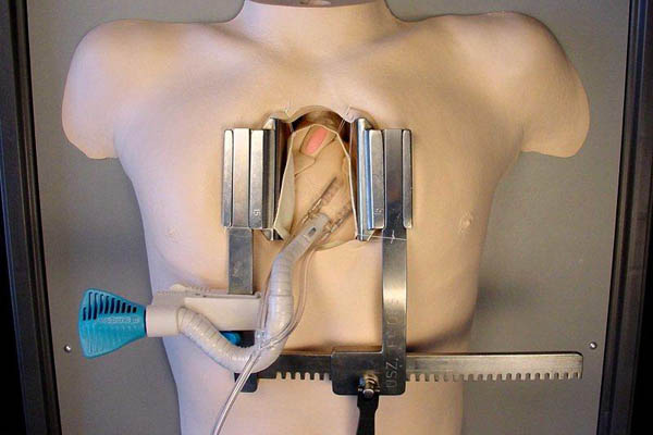
Much more effective are interactive computer programs featuring practical exercises and even virtual autopsies on the screen. Physiology can be experienced through harmless self-study on one’s own body. Various types of myography can be used instead of frog muscles to measure the electrical activity of nerves and muscles in a student’s thumb. Synthetic models for surgical training are well suited to develop and refine one’s skills. To study an animal’s anatomy, it is not necessary to kill animals. Animals euthanized for medical reasons or animals found dead can be used for this purpose. Through plastination, i.e., the transition into a plastic-like condition, such animals are everlasting.
Future physicians learn conducting surgery by practicing on dead humans, and veterinarians use dead animals who died a natural death or were euthanized for medical reasons. In a next step, the students will assist an experienced surgeon until they are capable of conducting surgeries themselves under observation. More on animal experiments in education and training >>
Simulators and Virtual Reality

In the veterinarian program there are similar dummies, e.g., the dog Alberta of the U.S. company SynDaver. Future veterinarians can train on Alberta all kinds of imaginable procedures, from intubation and castration to tumor removal and bone surgery.
Virtual Reality for surgeons works like flight simulation in pilot training. This real time simulation includes video recordings from real surgeries and enables haptic perceptions, i.e., the surgeons feel when the instruments touch or cut tissue, or when the tweezers pull or push. The simulation program translates the sense of touch into the corresponding images on the screen drawn from a huge video database. More about this >>
Unparalleled benefits of animal-free research
Scientifically well developed in vitro test systems offer a number of clear advantages compared to animal experiments.
- Reliability
Models based on human cells and data produce clear results that can easily be replicated, which is not possible in animal experiments due to physiological and anatomical differences between humans and animals, among other factors. - Sensitivity
In vitro test systems respond in part much more sensitively to toxic influences than living animals. - Costs
Once they are established, in vitro test systems are significantly cheaper than animal experiments. - Duration
Studies on in vitro systems produce results within hours while studies including animal experiments may take weeks, months, or even years. - High throughput of substances
In vitro systems enable, for instance, toxicological studies that allow to study a big number of pharmaceuticals or chemicals simultaneously, while the number of possibilities is limited in systems using animal experiments.
Conclusion
Animal-free methods are much more than a mere “alternative” to animal experiments. The wide range of human-based, animal-free research methods makes it possible to gain reliable findings. This not only spares animals a gruesome and unnecessary death in the lab, but it also provides us humans with viable options in biomedical research. We urgently need to abolish the system of animal experiments in order to make way for such well-thought-out, animal-free methods to not keep blocking medical progress.
15/02/2021
Silke Strittmatter, biologist and Dr Corina Gericke D.V.M.
Further information
NAT Database - the database on animal-free research methods >>
Leaflet „Modern science approaches“
Order in our online shop >> or download as PDF for free >>
Video "Mini organs and multi-organ-chips - Scientific progress without animal experiments"
privacy policy.
References
- Sicherer als Tierversuche? Informationsdienst Wissenschaft, 20.08.2008
- Nervennetz auf dem Biochip statt Tierversuche, du und das tier, 2/2010, S. 18
- Lunge auf dem Chip, Welt der Physik, 25.06.2010
- Benam KH, Novak R, Nawroth J, et al.: Matched-comparative modeling of normal and diseased human airway responses using a microengineered breathing lung chip. Cell Systems 2016; 3(5): 456-466.e4
- Hassell BA, Goyal G, Lee E, et al.: Human organ chip models recapitulate orthotopic lung cancer growth, therapeutic responses, and tumor dormancy in vitro. Cell Rep. 2017; 21(2): 508-516
- Fachhochschule in Jena entwickelt „das Labor“ auf dem Chip, Thüringer Allgemeine, 21.07.2010
- Versuchskaninchen aus Silizium, Technology Review, Juli 2004, S. 45-48
- Organ-Chips sollen Tierversuche ersetzen. Spektrum.de, 03.08.2015
- Van den Brink FTG, Wigger T, Ma L, et al.: Oxidation and adduct formation of xenobiotics in a microfluidic electrochemical cell with boron doped diamond electrodes and an integrated passive gradient rotation mixer. Lab Chip 2016; 16(20): 3990-4001
- Kim S, LesherPerez SC, Kim BC, et al.: Pharmacokinetic profile that reduces nephrotoxicity of gentamicin in a perfused kidney-on-a-chip. Biofabrication 2016; 8(1): 015021
- Günter A. et al.: A microfluidic platform for probing small artery structure and function. Lab Chip 2010; 10(18): 2341-2349
- Wyss Press release: Harvard's Wyss Institute creates living human gut-on-a-chip, 27.03.2012
- Zietek T et al.: Intestinal organoids for assessing nutrient transport, sensing and incretin secretion. Scientific Reports 2015; 5: 16831
- Ortega-Prieto AM, Skelton JK, Wai SN, et al.: 3D microfluidic liver cultures as a physiological preclinical tool for hepatitis B virus infection. Nat Commun. 2018; 9(1): 682. doi: 10.1038/s41467-018-02969-8
- Blundell C, Tess ER, Schanzer AS, et al.: A microphysiological model of the human placental barrier. Lab Chip 2016: 16; 3065-3073. doi: 10.1039/c6lc00259e
- Nesmith AP, Wagner MA, Pasqualini FS, et al.: A human in vitro model of Duchenne muscular dystrophy muscle formation and contractility. J Cell Biol. 2016; 215(1): 47-56
- Lind JU, Busbee TA, Valentine AD, et al.: Instrumented cardiac microphysiological devices via multimaterial three-dimensional printing. Nat Mater. 2017; 3: 303-308
- Kwon D: Organs on chips. The Scientist, 28.08.2017
- Hamilton J: Minibrains could help drug discovery for Zika and for Alzheimer’s. NRP24, 13.11.2016
- 1964 Surgeon General‘s Report on Smoking and Health, National Center for Chronic Disease Prevention and Health Promotion
- Langley G: A regulatory Smokescreen. BUAV/ECEAE (Hrsg.) 2004, S. 9-10
- Johns Hopkins Bloomberg School of Public Health: Database analysis more reliable than animal testing for toxic chemicals. News Release, 11.07.2018