Animal testing and animal-free methods in surgery courses
Although living in the 21st century, it is a sad reality that animals or parts of dead animals are still being used in surgical training and residency programs. Large animals in particular, such as pigs or sheep, are anaesthetized, used as training objects, and killed at the end of the training session. An intolerable status quo, especially since humane and human-relevant methods are available. With this campaign, Doctors Against Animal Experiments (DAAE) wants to draw attention to this scandalous practice and encourage course providers to stop using animals.
Animal experiments are carried out for basic research in the surgical domain as well as for the development of new instruments or implants, and to test their compatibility. Practicing surgical skills on anesthetized animals: What sounds like a relic from old times is common practice in many surgery courses. Anyone who thinks that this is absurd is absolutely right since humans and pigs or humans and sheep are anatomically different. A few selected points of analogy seem to be enough for this barbaric practice to stick around.
This is particularly incomprehensible because of the heaps of non-animal methods that have been available for many years. They can depict the anatomical conditions of a human and realistically represent the most diverse variations of the anatomy of a healthy person as well as different manifestations of clinical pathologies. With regard to patient safety, it is unacceptable and negligent to allow future surgeons to be trained in the wrong system. In many emergency situations, quick and safe action is required. In order to provide these skills, a surgeon must be familiar with the anatomical structures and uniqueness of the human body and organism, and recognize them within seconds.
Why do these courses exist?
There are many medical disciplines that involve surgical tasks and therefore require surgical skills, such as vascular surgery, thoracic surgery, aesthetic surgery, orthopaedics, trauma surgery and many more. In order to learn the relevant skills that are required for operating, these must be practiced extensively by the future specialists so that the movements are also safely mastered when they assist in surgery or carry out their first surgery on a real patient.
Specialists who want to refresh certain skills or acquire new ones also need practice and training.
For this purpose, many university hospitals, and also some private providers and medical professional associations offer training courses. These can last from a few hours to several days and also take place in the context of other events such as annual meetings or trade fairs.
Medical courses including animal use are not limited to surgery: In some cases, animals are also used in courses in anaesthesiology or in emergency treatment of seriously injured people, as well as in product training courses, which are sometimes financed by manufacturers of medical devices.
Sequence of training courses
Pigs are most commonly used for surgical exercises, although their anatomy differs significantly from the human anatomy. (Source: Prall M. et al, Physica Medica 2017, 43)
Depending on the topic, only medical professionals may participate. First, there is a general introduction to the topic, then a specialist lecture on the skills to be learned in this course, then the upcoming surgery to be performed is presented and discussed.
Unfortunately, animals are still brought in especially for these courses. In many course descriptions that contain exact schedules for the respective days, the system reveals itself on closer inspection: there is often an item on the agenda called "Introduction to the anatomy of pigs/sheep" (1). This is not surprising: Although these courses are only attended by specialists, some of them with years of professional experience, their knowledge is based on human anatomy – the only anatomy that is relevant to their profession. The animal anatomy is very different from the human one. Therefore these differences must first be explained in detail before the exercise can start – which makes the scientific madness of using animals as models for humans more than clear. The skills are learned in the wrong system, which is negligent, especially with regard to patient’s safety, which must always be the main concern.
If animals are used in a course, they are first anaesthetized and strapped to the surgery table. Then, the surgery on the animal begins, often leading to complications. In a study in which 40 pigs had a pulmonary lobe removed, more than 50% developed impaired breathing, resulting in a 10% death rate in the last trimester of surgery. 20% of the animals developed an extremely slow heart rate, resulting in 5% of the pigs dying before the end of the procedure (2).
After the end of the operation, the animals – if they have survived that far – are usually killed while they are still under anaesthetic.
Where do these courses take place?
Throughout Germany, there are courses that use animals in advanced or postgraduate training, as our map shows. The providers are very diverse: surgical departments of (university) hospitals, professional associations of certain medical specialties, but also medical equipment manufacturers can be the organizers. In addition, there are specialized centres that offer multidisciplinary courses throughout the year.
However, many organizers do not clearly communicate on their websites and flyers whether animals or parts of dead animals are used in the training or whether animal-free models are used.

Animal use in surgical training and advanced education courses. Results of research by Doctors Against Animal Experiments. The map does not claim completion.
The oppose the reigning non-transparency, we have compiled an overview of the surgical courses that are offered in Germany. It is based on internet research, the help of our physician members, who gave us indispensable input, and direct inquiries to the providers. In the list of 59 course providers, 19 definitely work without animals or animal organs; they use one or more simulation methods. Twenty-three providers use animals, and for 17 providers, it could not be clearly ruled out that animals were used in these courses. The communication with the organizers could not shed light on every case, as about half of the institutions addressed did not respond even to multiple requests, or the responses were so ambiguous that animal use could not be ruled out with certainty. This non-transparency, along with the fact that inquiries of this kind are often being ignored, raises the suspicion that animal protection is not highly valued in these institutions.
Accordingly, we have once again written to all providers, criticizing those whose courses involve animal use and demanded a switch to human-based methods.
While it is gratifying that about a third of the providers work with animal-free methods, overall, it is distressing that animals are still used in so many other courses. Above all, this raises the question which (legal) arguments are made to justify the use of animals when other providers are able to provide animal-free courses.
Figures and legal basis
The number of animals used and killed for education has been at a similar level for years and has only slightly decreased over the past 10 years. In 2019, according to statistics from the German Federal Ministry of Agriculture (BMEL), 53.805 animals were used for this purpose, including 1.504 pigs and 108 sheep (3). No further details are provided, so it is unclear how many animals are killed specifically for surgical training. Although the number may seem low compared to the total number of animals used in animal experiments, each individual animal is subjected to significant suffering during its short life. In addition, learning surgical techniques in the wrong species poses a risk to the health of human patients – even though these courses are supposed to save their lives.
From a legal point of view, animal experiments for education, training, and advanced education are permitted according to § 7a paragraph 1 sentence 7 of the Animal Protection Act, given the fact that they are indispensable (4).
It is questionable, of course, to what extent one can speak of indispensability, when numerous animal-free methods exist. The law states "[...] It must be examined whether the purpose pursued cannot be achieved by other methods or procedures. [...]" (4). However, this is the crux of the matter: animal experiments for education, training, and advanced education purposes were previously only subject to a so-called obligation to notification under the German Animal Protection Act, i.e. a formally correctly completed form was sufficient to be able to carry out such experiments. It sufficed for the applicants to state that no adequate animal-free model was available. An independent review was not required until now, and a rejection of the notification, thus, virtually impossible.
It is scandalous that this practice, which is enshrined in German law, had violated applicable EU law for 8 years, since the EU Animal Experiments Directive stipulates that animal experiments for educational purposes must be subject to (active) approval (5). With the infringement decision of the European Commission against Germany for incorrect implementation of EU law, the German government was forced to alter the requirements for applications for experiments for education, training, and advanced education. Whether this regulation will affect the practice of surgical training remains to be seen (6).
The fact that politicians have shown little interest in protecting animals in the long term is also shown by a so-called minor inquiry in 2013 to the Berlin Senate. When asked to what extent the Senate can exert influence to ensure that a well-known course provider uses modern non-animal training methods instead of animals, the answer was: "Since these are legally tested and approved as well as medically valuable training courses and research projects in the sense of animal protection, the Senate sees no need for action in this regard." (7) This clearly shows a lack of genuine commitment. Just as in so many other cases, considerable political amendments are necessary in this area.
Differences between humans and animals
The similarities between humans and animals are mentioned over and over again by people who support animal testing. Differences are rarely discussed, although they cannot and should not be dismissed.
Pigs are used in thoracic surgery particularly often, although the literature generally rates them as a poor model for humans (8). Other publications specifically refer to their short and narrow chest (2) compared to humans. The heart of a pig has a different shape than that of humans, and there are also some noteworthy physiological differences: the left atrium of the pig has only two veins, while there are four veins in humans. A conspicuous muscle band is localized in the pig's right ventricle at a much higher position than in humans (9).
Because such traits can serve as points of orientation during surgery, there is a high risk that they will be learned incorrectly if studied in the wrong species. Furthermore, the distribution and size of the renal-arterial segments are also completely different in pigs (10). In general, the human aorta differs from that of animals in vessel geometry, size, and physiological conditions (11). And those are just a few examples of the considerable anatomical differences between humans and pigs.
Sheep are said to be more similar to humans when it comes to the pulmonary system, but there are also several differences noted in the literature. The right lung in particular differs from that of humans: the vena cava is larger, and the arterial plexus is located behind it. Moreover, the upper pulmonary lobe has its own access to the trachea (8).
But even without these differences, there would be no reason to use animals as surgical models. There are countless methods and models that accurately represent the human anatomy and with which novice and experienced surgeons can learn and train in the correct species.
Useful surgical training methods
Human cadavers
These are the bodies of voluntary human donors who consented during their lifetime to donate their bodies to science after their natural death. There are different ways of preserving bodies. The most suitable method is probably the Thiel procedure, in which the natural colour, elasticity, and properties of the individual tissues are retained, and the surgical experience is as authentic as possible (12). The big advantage of the Thiel procedure is that it makes it possible to train almost every surgical procedure of any specialty.
To enhance reality, the body can be connected to a pump so that blood flow and pressure are generated artificially but realistically. Both can also be varied to simulate different physical or pathological conditions. Furthermore, temperature is adjustable. Endovascular training can thus be realistically performed, and endovascular medical devices can even be tested realistically in an anatomically correct system. Many procedures are performed under fluorescent dye imaging, and this method is also very well suited for this purpose (11).
All accessories to turn the donated corpses into a surgical model can be purchased commercially as a kit. Blood solutions that correctly mimic blood clotting and change the colour of the blood, depending on environmental conditions of the tissues surrounding, are also available. The body can even be reused several times over a period of 2 weeks. It is also possible to practice amputations with these models (13).
Physical models
These are dummies that provide an anatomically correct reproduction of the human body or certain parts of it. Different materials are used to recreate a realistic experience of the various tissues of the human body.
The TraumaMan is well-known. It is used for emergency training (14), i.e., in simulated accidents where very fast action of the entire emergency response team is required. Various complications can be set in advance. In addition, artificial blood circulation can be generated so that realistic bleeding can occur in the event of cuts or errors. The modular design of head, upper and lower body makes it possible to use the TraumaMan as a whole or in certain body sections. Interchangeable materials offer each trainee the possibility of performing each intervention by themselves. In the USA, the dummy has been used for ATLS training (Advanced Trauma Life Support Training) since 2001.
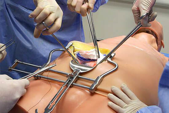
Upper body of the TraumaMan (©Skills Med Deutschland GmbH)
Some providers have specialized in certain medical fields. For example, the company Gaumard offers a wide range of simulation models for obstetrics, neonatology, and pediatrics. (15)
Especially for training medical professionals to be prepared for working in crisis areas (i.e., during a war), there are devices such as the "Cut Suit," which realistically recreates war scenarios on people. It is a kind of artificial, somewhat thinner torso that containing dummies of the relevant organs and fake blood. The suit is strapped on an actor and worn around the (actual) body. In this way, the emergency situations such as blood loss and organ injuries can be simulated realistically and trained appropriately (16).
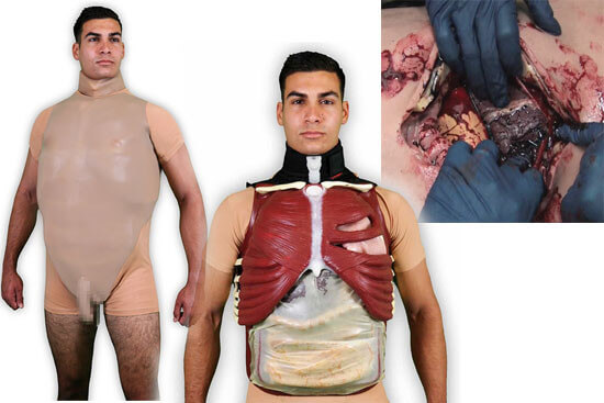
The Cut Suit can be used to recreate realistic war injuries (©www.strategic-operations.com)
The U.S. company SynDaver offers a lifelike, full-size human "doll" that is intricately handcrafted from a wide variety of materials. Many of the materials are developed and manufactured in-house to create a resemblance as close as possible to human tissue.
The models include a muscular, skeletal, cardiac, and vascular system, as well as fascial tissue and many elements of the nervous system. The respiratory tract, gastrointestinal tract, and urogenital tract are also present. The training of various surgeries is possible, such as intestinal resection (partial removal of the intestine), appendectomy, angioplasty (widening of a narrowed blood vessel), and the placement of stents with the help of fluoroscopy. Limbs and various internal organs are also available individually (17).
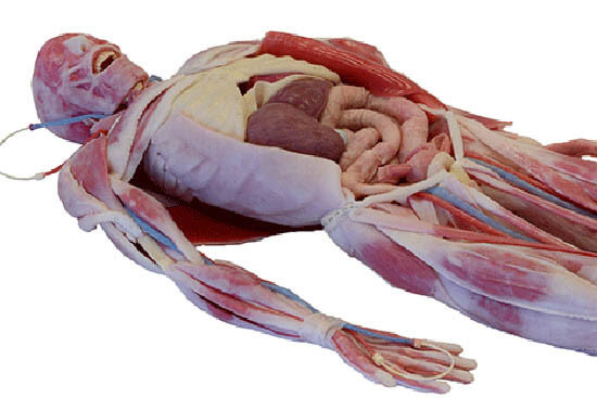
Life-like model made by SynDaver (©SynDaver)
3D printing can be used to produce patient-specific body parts such as hearts from CT scan templates, on which basic surgeries can be planned better. The entire bone system as well as vascular models can be printed as models in healthy or pathological form. (18) (19).
The "Hamburg Anatomical Neurointerventional Simulation Model" (HANNES) is already successfully implementing this: The treatment of aneurysms (protrusions in blood vessels that can become life-threatening) in advanced education is already being trained on patient-specific 3D printed models. This allows physicians to repeat various pathological cases as often as they wish (20).
Virtual simulations
This principle, which is similar to flight simulators used for pilot training, is implemented using computer programs. Advances in virtual technology allow very realistic representations that can hardly be distinguished from real surgical videos. The latter can be integrated into some virtual modules.
This area is particularly suitable for training minimally invasive techniques. The entire range of these surgeries can be demonstrated particularly well, since this surgical technique does not involve large incisions in the abdomen, thorax or joint to reach the actual operation site. Instead, only several small incisions are made at strategically useful points on the body. Through these openings, instruments specifically suited for these surgeries as well as a camera with light function are inserted into the body. The surgeon follows and orients himself via the camera's image transmission and performs the surgery "on the screen". Most surgeries are now planned using these procedures as they are less invasive and therefore increase the patient's chances of survival, as well as significantly shortening the recovery period in some cases (21).
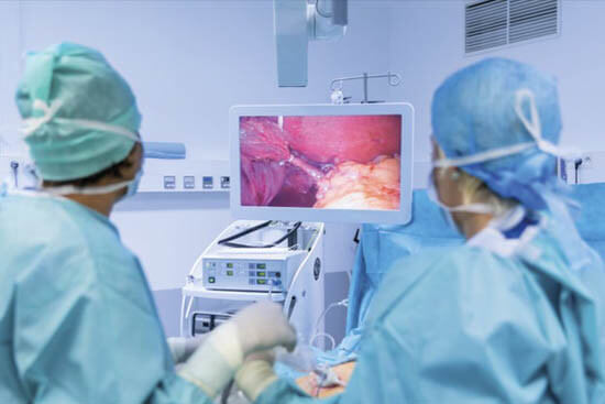
Virtual simulation can be used to practice operations in minimally invasive surgeries.
These systems often look like a box with a screen, just like the one by SurgicalScience, and are sometimes even compact enough to be transportable. Different programs can be set. Beginners can first practice basic techniques such as suturing and clamping. For more advanced exercises, original surgical videos are also available, so that the respective procedure can first just be watched before the user's own practice course starts, which can optionally be accompanied by interactive instructions. Modules for gall bladder surgery, lung lobectomy, uterus removal, hernia surgery, and many other procedures are available. A big advantage is that the supervisor doesn’t have to be constantly present. The course participants can practice techniques independently and at their own pace until they have mastered them with confidence (22).
These systems are even available for certain basic techniques as a “to go” version and can be connected to a smartphone, which makes it possible to do dexterity exercises during business trips, for example (23).
These virtual modules can also be combined with a physical model. The image below depicts a part of the body covered with synthetic materials, for example a knee, into which surgical instruments can be inserted and the actual surgery can be performed and practiced virtually on the screen using a corresponding computer module (24).
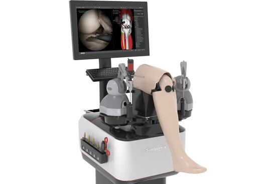
Arthro Knee for practicing knee operations (©Simbionix ARTHRO Mentor, 3D Systems)
What is so special about these virtual models and the surgical exercises they enable are the realistic haptics - different tissues show different levels of resistance to the instruments. Those are implemented by the programs as they would behave in a real invasive procedure. In very soft tissues, which are delicate and can be injured quickly, the program allows the instruments to penetrate faster accordingly. Firmer structures require more force.
The difficulty level of the surgeries can also be adapted to the learning level. Since every human body is unique, there are many different anatomical conditions, both of healthy and pathologically altered tissues and structures. These show the range of variety for which physicians must be prepared.
Nordic Simulators offers customized, virtual 3D scenarios that take place around an actual emergency procedure on a human model: Crisis situations such as pileups can be visually depicted on a curved video screen; a sound system gives a realistic impression of the situation and reinforces the realistic setting. Physicians and medical staff are exposed to extreme stress in such cases, so safe actions can be trained even in very challenging situations (25).
Virtual dissection tables might be less relevant for surgical courses but are very interesting as an alternative to dissections of donor bodies for medical students. These are visually based on operating tables, have a touch-screen interface, and offer a wide variety of levels that can also be zoomed into. Over 1,000 pathological cases provide comprehensive teaching material. A radiology module offers additional imaging procedures (26).
There is also anatomy software for home use, some of which is even free of charge (27,28). The apps “Human Body Atlas” and “Complete Anatomy” offer the possibility to work on surgical skills on a smartphone, on the bus or train for example.
P.O.P. trainer
The P.O.P. (= Pulsatile Organ Perfusion) trainer is a type of plastic box containing a single organ, which can be perfused with artificial blood by a pump and into which instruments are inserted through openings. In this way, necessary skills for minimally invasive surgery ("keyhole surgery"), can be learned. In those procedures, instruments and a camera are inserted into the body through natural body openings or small incisions, and the devices are controlled via a screen.
Note: The organs in question are animal organs, mostly slaughterhouse waste. Thus, animals are not killed specifically for this purpose, but the anatomical differences between humans and animals are still a scientific obstacle, of course. Some might argue that this way, organs from animals that were going to be killed anyway are going to good use. Considering the aforementioned crucial differences between humans and animals, this “good use” is questionable in and of itself. Furthermore, it has to be considered that this surplus in animal organs only exists due to the excessive demand for meat as “food”. The use of animal organs in surgical training is therefore neither scientific nor ethical.
However, it is also possible to equip the box with human models made of various synthetic materials. There are various organs that can be used several times, such as the entire intestinal tract or a construct on which the removal of the gallbladder can be practiced (31).
Due to the correct anatomical points of orientation, this is a human-relevant and, with the use of synthetic or human material, an ethically justifiable training method.
There are way more human-relevant, ethically sound, and scientifically sensible training methods. The NAT database www.nat-database.org/ provides an overview.
Better learning success - human-relevant, ethically flawless
Numerous publications show that the learning success with non-animal models is at least equivalent, often even better than if based on animal models. Of course, it is also important to identify the right model for the particular procedure to be learned. Virtual models are more appropriate for airway procedures (8), while course participants found human bodies to be an ideal training model for urologic surgeries (32,33). Those are also the best option when it comes to learning so-called open surgeries that require, for example, opening the chest (21).
Lots of publications compare human-relevant methods with animal experiments. Although some advantages of animal-based methods are mentioned (such as realistic surgical situation and similar anatomy), the limitations follow on the spot: similarities with human anatomy are often isolated and limited to certain areas. Furthermore, they are associated with high costs for preparation and raise ethical concerns. In addition, a supervisor must always oversee the exercises, and many procedures can only be performed once. This is particularly significant because it is estimated that trainee surgeons have to perform at least 100 individual exercises to learn basic skills (8).
The alleged limitations of animal-free systems, which are described in the literature, mostly relate to costs or the lack of certain software modules, some of which are still considered to be expandable. However, these are not obstacles that can and should prevent us from using non-animal methods in surgical courses. Software modules can be created in a manageable amount of time and the more frequently simulators are used, the cheaper they become.
Some providers actually justify the use of animal models with scarce time and financial resources (34), which is a clear violation of the Animal Welfare Act, which states: "[...] pain, suffering or harm may only be inflicted on animals to the extent that it is essential for the purpose pursued; in particular, they may not be added for reasons of labour, time or cost savings. [...]" (4)
Wherever your focus may lie: There is no scientific, no medical, and certainly no ethical reason to keep using animals in surgical training. On the contrary - only advantages can be expected from a shift to animal free methods.
The situation in the USA shows that none of this is utopian: three quarters of the surgical programs are already animal-free – and counting! Throughout their whole university education, medical students are now learning completely without animal use. This was achieved primarily through the work of the Physicians Committee for Responsible Medicine (PCRM), which has been doing intensive educational work in this regard for decades (35).
This can serve as a role model for our campaign to make course providers aware of the fact that the use of human-relevant training methods is the first and best option for surgery - and DAAE’s also applies to this case: in the interest of humans and animals.
29 March 2021
Dipl. Biol. Julia Radzwill
References
- Deutsche Gesellschaft für Thoraxchirurgie. 13. Operationskurs -Techniken der fortgeschrittenen Thoraxchirurgie. 2020
- Tedde ML, Brito Filho F et al. Video-assisted thoracoscopic surgery in swine: an animal model for thoracoscopic lobectomy training. Interact Cardiovasc Thorac Surg. 2015; 21(2):224–30
- Bundesministerium für Ernährung und Landwirtschaft (BMEL). Versuchstierdaten 2019
- Bundesministerium für Justiz und Verbraucherschutz. Tierschutzgesetz. Fassung vom 18.05.2006, zuletzt geändert am 19.06.2020
- Bund für Risikobewertung. Richtline 2010/63/EU zum Schutz der für wissenschaftliche Zwecke verwendete Tiere.
- BMEL Pressemitteilung. Mehr Schutz für Versuchstiere. 20.01.2021
- Senat von Berlin. Kleine Anfrage Bündnis 90/Die Grünen 17/12695 vom 26.09.2013
- Haidari T, Konge L. et al. Simulation for the video-assisted thoracic surgery surgeon. Video-Assisted Thoracic Surgery 2019; 4(12)
- Crick SJ, Sheppard MN et al. Anatomy of the pig heart: comparisons with normal human cardiac structure. J Anat 1998; 193(1);105–19
- Pereira-Sampaio M, Favorito LA et al. Proportional analysis of pig kidney arterial segments: differences from the human kidney. J Endourol. 2007; 21(7):784–8
- McLeod H, Cox BF et al. Human Thiel-Embalmed Cadaveric Aortic Model with Perfusion for Endovascular Intervention Training and Medical Device Evaluation. Cardiovasc Intervent Radiol. 2017; 40(9):1454–60
- Thiel W. The preservation of the whole corpse with natural color. Ann Anat. 1992; 174(3):185–95
- Maximum Fidelity Surgical Simulations. Hyper-realistic surgical simulator.
- Simulab Corporation. TraumaMan Surgical Simulator
- Simulators for Health Care Education
- Strategic Operations. Medical Simulation Products
- SynDaver Surgical Model
- Stratasys Direct. Anatomical Model 3D Print
- OS Implants. Anatomical Model
- Informationsdienst Wissenschaft. Pressemitteilung. Alternative zum Tierversuch: TUHH-Forschende entwickeln neues Trainingsmodell für Aneurysmabehandlungen. 20.01.2020
- Konge L, Petersen RH, Ringsted C. Developing competency in video-assisted thoracic surgery (VATS) lobectomy. J Thorac Dis. 2018; 10(17):S2025–8
- Healthcare LAP-brochure. 2020
- Evidence based training.
- ARTHRO Mentor Broschüre 2019
- Nordic Simulators. Simulation Centers Design & Consulting
- Anatomage Table Deutschland
- Zygote Body 3D Anatomy Online Visualizer. Human Anatomy 3D
- Interactive 3D Anatomy - Disease Platform
- Visible Body App. Virtuelle Anatomie für Einblicke in den menschlichen Körper
- 3D4Medical App. 3D Human Body Atlas
- Simulab Corporation. Laparoscopic Trainers
- Healy SE, Rai BP et al. Thiel embalming method for cadaver preservation: a review of new training model for urologic skills training. 2015; 85(3):499–504
- Soler-Silva Á, Sanchís-López A et al. The Thiel cadaveric model for pelvic floor surgery: Best rated in transferable simulation-based training for postgraduate studies. Eur J Obstet Gynecol Reprod Biol. 2021; 256:165–71
- Rudolf-Zenker-Institut für Experimentelle Chirurgie. Viszeralchirurgie Kompakt. März 2020
- Physicians Committee for Responsible Medicine. Surgery Training